


See letter "Comment on “bronchus perforation by EZ-BlockerTM endobronchial blocker during esophageal resection after neoadjuvant chemoradiation”" in Volume 72 on page 616.
/W3C//DTD HTML 4.01 Transitional//EN" "http://www.w3.org/TR/html4/loose.dtd">
Bronchus perforation by EZ-BlockerTM endobronchial blocker during esophageal resection after neoadjuvant chemoradiation -a case report-
-
Jorien M van de Pas1
 , Margaretha CE van der Woude2,3, Henricus J Belgers1, Karel WE Hulsewé1, Erik R de Loos1
- Received August 14, 2018 Revised December 13, 2018 Accepted December 14, 2018
- Abstract
-
- Background
- Double-lumen tubes (DLT) and endobronchial blockers (EB) are used for one-lung ventilation in thoracic surgery. More complications are seen when using DLT when compared to EB, while major complications are rarely seen.
- Case
- This case report describes a perforation of the right mainstem bronchus by an EZ-Blocker EB in a patient undergoing a minimally invasive esophagectomy after neoadjuvant chemoradiation.
- Conclusions
- We advise to insert an EZ-BlockerTM EB with caution and only under direct bronchoscopic visualization, especially in previous irradiated patients.
One-lung ventilation (OLV) is a challenging anesthetic technique used in thoracic surgery, including minimally invasive transthoracic esophagectomy. Single-lumen tubes (SLT) with endobronchial blockers (EB), or double-lumen tubes (DLT), are routinely used for the selective blockage of one lung to facilitate OLV. The first DLT was introduced in 1949, and has for a long time been the most commonly used device for this technique [1]. It consists of an endotracheal lumen terminating in the trachea and an endobronchial lumen positioned in the mainstem bronchus, allowing independent OLV (Fig. 1). An alternative device for lung isolation is a endobronchial blocker, a balloon-tipped semi-rigid catheter that is inserted through a standard SLT. Numerous types of EB have been developed during the last few decades, among which is the EZ-BlockerTM (RuschⓇ, Anaesthet-IQ, the Netherlands), a new type of EB with a Y-shaped distal end that mirrors the carina, and contains a cuff on each side that can be inflated for selective bronchus blocking (Fig. 2). The EZ-BlockerTM is advanced through the trachea and placed in the carina under direct bronchoscopic guidance [2–4]. Common complaints with the use of both techniques are hoarseness and sore throat; major complications like airway rupture are very rare, but have also been reported [2,4,5].
This case report describes a mainstem bronchus perforation after placement of an EZ-BlockerTM for OLV during a minimally invasive transthoracic esophagectomy. As far as the authors are aware, this is the first description of a bronchial perforation using an EB.
- Case Report
- Case Report
A 78-year-old male with a history of coronary artery disease, angina pectoris, and hypercholesterolemia was diagnosed with a cT3N2M0 adenocarcinoma of the distal esophagus. After neoadjuvant chemoradiation (41.4 Gy in 23 fractions with 5 weeks of Carboplatin and Paclitaxel), a minimally invasive transthoracic esophagectomy according to Ivor Lewis was scheduled. A preoperative PET-CT scan showed no detectable change in the mainstem bronchus.The operation was performed under general anesthesia with antibiotic prophylaxis. To achieve OLV, an SLT was used in combination with an EZ-BlockerTM EB. Before insertion, the head of the patient was placed in the midline position. The endotracheal tube was withdrawn under direct vision using fiberoptic bronchoscopy until it was 4 cm above the carina. After connecting the multiport adapter, the EZ-BlockerTM EB was advanced with deflated cuffs and positioned at the carina under direct bronchoscopic guidance. The right cuff was inflated, deflated, and marked. The volume of air required to inflate the cuff was noted.During the laparoscopic phase in the supine position, a standard lymph node dissection was performed. Next, the gastric conduit was created and a feeding jejunostomy tube was placed. The patient was then placed in the prone position for the thoracoscopic phase of the operation. While turning the patient, the cuffs of the EZ-BlockerTM EB were kept deflated to reduce the risk of traumatic complications. The position of the EZ-BlockerTM EB was rechecked after turning the patient, but bronchoscopic inspection was more difficult with the patient in the prone position because of an excessive central airway collapse below the level of the tube. At the start of the thoracoscopic stage of the surgery, two-lung ventilation was supplied with pressure-controlled ventilation using low driving pressures, with the following ventilator settings: tidal volume (Vt), 300 ml; respiratory rate (RR), 20 /min; peak pressure (Pmax), 14 cmH2O; positive end-expiratory pressure (PEEP), 3 cmH2O; and fraction of inspired oxygen (FiO2), 0.85. The patient was hemodynamically stable. A pneumothorax was created by insufflation of carbon dioxide at a maximal pressure of 7 mmHg. The inferior pulmonary ligament was divided. After opening the pleura towards the right mainstem bronchus, the tip of the EB unexpectedly came into view, perforating the bronchus (Fig. 3). The esophageal dissection was completed and the resection specimen was stapled to the gastric conduit; the proximal esophagus was transected near the thoracic inlet.A bronchoscopy was performed via the endotracheal tube. Both distal extensions of the EB appeared to be in the right mainstem bronchus. The left arm of the Y-shaped tip was perforating the bronchus 4–5 cm distal to the carina, and the right arm was positioned more distal, intraluminal in the ostium of the right lower lobe bronchus.The SLT was exchanged for a 39 French left-sided DLT with the perforating EB still in place. The patient was positioned in the left lateral decubitus position. The right mainstem bronchus was selectively blocked. The ventilator settings were: Vt, 240 ml; RR, 16 /min; Pmax, 21 cmH2O; PEEP, 8; FiO2, 0.80. The patient remained hemodynamically stable. A right posterolateral muscle-sparing thoracotomy was performed. The mediastinal pleura was opened near the carina, after which the EZ-BlockerTM EB was identified perforating the pars membranacea on the dorsal aspect of the right mainstem bronchus (Fig. 4). The perforating EB was removed by the anesthesiologist under direct vision. The resulting bronchial defect of approximately 2 mm was closed primarily using interrupted 3-0 polydioxanone (PDSⓇ, Ethicon, Johnson & Johnson, the Netherlands) sutures. Air tightness was confirmed with an underwater test. The closed defect was covered with an intercostal muscle flap for extra protection of the vulnerable irradiated tissue. To diminish the risk of postoperative complications, no anastomosis was made. An end cervical left-sided esophagostomy was performed with the intention of reconstructing the gastric conduit at a later stage. The patient was extubated directly post-surgery.A backup plan in the event of a respiratory insufficiency consisted of selective left mainstem bronchial intubation, followed by OLV of the left lung with lung-protective ventilation using low tidal volumes, higher PEEP levels, and low driving pressures. The patient recovered well and was discharged on day 14.A follow-up PET scan 2.5 months after surgery unfortunately showed adrenal and chest wall metastases. After consulting the patient and family, it was decided to start palliative radiation therapy and to renounce further surgical procedures.
- Discussion
- Discussion
Iatrogenic mainstem bronchus perforation caused by an EZ-BlockerTM EB is a rare complication and, as far as the authors are aware, has never been described in the literature.Both DLTs and SLTs with EBs are widely used for OLV. The DLT is more rigid and has a larger diameter compared to the SLT with an EB [4]. The disadvantages of the DLT include challenging placement in difficult airway scenarios, inadequate sealing when inappropriately sized, the need for tube exchange when prolonged ventilation is required, and the risk of tracheobronchial injury, or even bronchial rupture [2,4,5]. Tracheobronchial rupture is caused by the larger DLT external diameter compared to the internal diameter of the bronchus, while mainstem bronchus rupture results from overinflation of the tracheal cuff. We found two reported cases of rupture of the left main bronchus after radiotherapy with use of a DLT [5,6], and using an SLT with an EB seems to be an interesting alternative. It is more flexible, easier to insert, and leads to less complications compared to using a DLT [2,4]. Moreover, no major complications have been described when using an SLT in conjunction with an EB.An EZ-BlockerTM is placed horizontally through, or together with, a single-lumen endotracheal tube with the patient in the prone position. The endotracheal tube must be fixed in the midline to prevent directing both cuffs into one of the mainstem bronchi. At least 4 cm should separate the distal end of the endotracheal tube and the carina, so that the Y-shaped distal end of the EZ-BlockerTM can deploy correctly. Resistance indicates correct placement of the EZ-BlockerTM at the carina, but bronchoscopy should still be used to confirm correct placement [2–4]. Mourisse et al. [2] describe malposition of the EZ-BlockerTM EB with both extensions placed in the right mainstem bronchus in 50% of the cases after blind insertion. Additionally, malposition occurs in 40% of the cases where the patient is turned to the lateral position, and in 8.3% of the cases during OLV [2]. No data are available for malposition in the prone position.This case report describes a perforation of the right mainstem bronchus by an EZ-BlockerTM EB in a patient undergoing minimally invasive esophagectomy following neoadjuvant chemoradiation. Despite careful introduction, a perforation occurred in vulnerable tissue. Regardless of this rare complication, the SLT with an EB remains our device of choice for OLV during thoracoscopic esophageal surgery because of the ease of use and low complication rate.We advise that an EZ-BlockerTM EB should be inserted with caution and only under direct bronchoscopic visualization, especially in previously irradiated patients. During time-out or briefing before the induction of anesthesia, it should be mandatory to take into consideration recent radiotherapy located close to central airways.
- Acknowledgments
Ernst van Loon, independent photography professional and clinical photographer at Zuyderland Medical Center – Figs. 1 and 2.
- Conflicts of Interest
No potential conflict of interest relevant to this article was reported.
- Notes
-
Author Contributions
Jorien M van de Pas (Writing – original draft; Writing – review & editing) Margaretha CE van der Woude (Writing – review & editing) Henricus J Belgers (Writing – review & editing) Karel WE Hulsewé (Conceptualization; Writing – review & editing) Erik R de Loos (Supervision; Writing – review & editing)
-
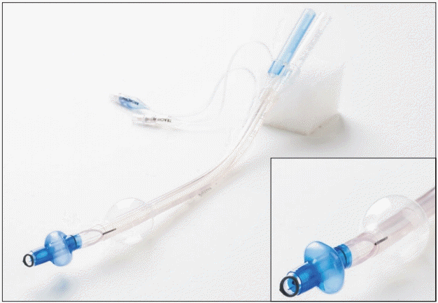
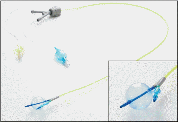
Fig. 3.
EZ-BlockerTM endobronchial blocker perforating the right mainstem bronchus (Video-Assisted Thoracic Surgery, VATS).

Fig. 4.
EZ-BlockerTM endobronchial blocker perforating the right mainstem bronchus dorsally (posterolateral muscle-sparing thoracotomy).

- References
- 1. Wang K, Atul C, Mehta J. Flexible Bronchoscopy. 3rd ed. Chichester, John Wiley & Sons. 2012, p 262.2. Mourisse J, Liesveld J, Verhagen A, van Rooij G, van der Heide S, Schuurbiers-Siebers O, et al. Efficiency, efficacy, and safety of EZ-blocker compared with left-sided double-lumen tube for one-lung ventilation. Anesthesiology 2013; 118: 550-61.
[Article] [PubMed]3. Verelst PL, van Zundert AA. Use of the EZ-Blocker for lung separation. J Clin Anesth 2013; 25: 161-2.
[Article] [PubMed]4. Ruetzler K, Grubhofer G, Schmid W, Papp D, Nabecker S, Hutschala D, et al. Randomized clinical trial comparing double-lumen tube and EZ-Blocker for single-lung ventilation. Br J Anaesth 2011; 106: 896-902.
[Article] [PubMed]