Combined supraclavicular and superficial cervical plexus block for clavicle surgery
Article information
Abstract
Background
Clavicle fractures occur in 35% of shoulder girdle fractures. Surgical fixation is preferred, especially in young patients for optimal functional outcomes, while nondisplaced fractures are usually treated conservatively.
Case
A 38-year-old male patient was admitted to the emergency services with a fracture of the left clavicle following a fall. During the preoperative evaluation, the patient requested to be awake during the surgery. Combined supraclavicular and superficial cervical plexus block was performed under ultrasound guidance without complications and the patient experienced no pain.
Conclusions
This technique may avoid possible complications related to interscalene brachial plexus block. Future studies are required to confirm the safety and efficacy of this approach.
Clavicle fractures occur in 35% of shoulder girdle fractures. While nondisplaced fractures are usually treated conservatively, surgical fixation is preferred especially in young patients for optimal functional outcomes [1]. Clavicular surgery can be performed under general or regional anesthesia with peripheral nerve blocks [2].
The supraclavicular nerves originate from the superficial cervical plexus and innervate the skin overlying the clavicle [1,3]. The superior trunk of the brachial plexus also includes the supraclavicular branches including the dorsal scapular nerve, the long thoracic nerve, the suprascapular nerves, and the nerve to the subclavius that arise superior to the clavicle [3]. Opinions vary regarding the innervation of the clavicle. Pain transmission may be mediated by the superficial cervical plexus or the subclavian nerve alone; however, the precise pathway is uncertain. The superficial cervical plexus block may be used alone or in combination with the deep cervical plexus block, interscalene brachial plexus block, or supraclavicular brachial plexus block for surgical anesthesia and postoperative analgesia for clavicle surgery [4]. When compared to the interscalene brachial plexus block, the supraclavicular brachial plexus block has lower rates of phrenic nerve palsy [5]. The supraclavicular brachial plexus block carries the risk of pneumothorax; however, it occurs only when the inferior divisions are blocked. A written informed consent was obtained from the patient prior to the procedure.
Case Report
A 38-year-old male patient was admitted to the emergency services after he had slipped and fallen on an icy footpath that resulted in a fracture of the left clavicle. A consultation was requested from Department of Orthopedics and Traumatology. Following evaluation by the orthopedic surgeon, open surgery with internal fixation was planned. The patient had no previous history of surgery, drug usage, or allergy and had no co-morbidities. During the preoperative evaluation, the patient requested to be awake during the surgery. A peripheral nerve block was planned and the patient was offered detailed information regarding the procedure.
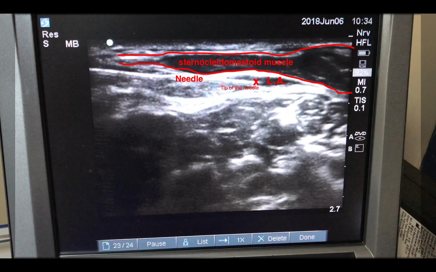
In the operating room, monitoring was established with electrocardiogram, SpO2 measurement, and non-invasive blood pressure measurement. An intravenous infusion of 0.9% sodium chloride was commenced. Midazolam, 2 mg, was administered intravenously for sedation. The patient was placed in a semi Fowler’s position, head rotated to right 30 degrees. The left side of the neck was cleaned with povidone-iodine. The block site and the surface of the ultrasound probe were covered with a sterile dressing. A linear transducer was placed at the level of the thyroid cartilage at the posterior border of the sternocleidomastoid muscle. The carotid artery was identified on scanning the neck in an anteroposterior direction. Color Doppler was used to confirm the presence of other blood vessels at the site of the block. The probe was moved laterally after identification of the carotid artery and the posterior border of the sternocleidomastoid muscle was centered on the screen. Using an in-plane technique, the needle was inserted close to the lateral end of the probe from lateral to medial through the thyroid cartilage. The tip of the needle was tracked under ultrasound guidance and positioned in the fascia deep to the sternocleidomastoid muscle. The local anesthetic solution was administered after confirmation of the needle tip position and a negative aspiration test. A mixture of 10 ml of 0.5% bupivacaine and 5 ml of 2% lidocaine was injected into the superficial cervical fascia (Fig. 1).

A full view of the needle and the spread of local anesthetic (L.A.) solution deep to the sternocleidomastoid muscle.
We decided to perform a supraclavicular brachial plexus block to avoid the side effects of the interscalene approach. The probe was placed in the supraclavicular fossa parallel to the clavicle. In contrast to the classic approach to supraclavicular brachial plexus block, we aimed to block the divisions originating from the superior trunk alone. After identifying the pulsations of the carotid artery, we applied color Doppler for identification and confirmation of any other vascular structures at the site of needle insertion. The needle was inserted using an in-plane technique from lateral to medial. The needle was tracked under vision and advanced through the platysma muscle and the fascia covering the brachial plexus. After the needle had passed through the fascia, a mixture of 5 ml of 0.5% bupivacaine and 5 ml of 2% lidocaine was injected after a negative aspiration test. After confirmation of the absence of pain, surgery was commenced. The procedure lasted for nearly 2 hours during which there was no significant variation in heart rate or blood pressure and the patient did not experience any pain. No complications such as Horner’s syndrome or phrenic nerve palsy were observed during the procedure; no additional opioids or benzodiazepines were administered (Fig. 2).
Discussion
We prefer regional anesthesia techniques for clavicular surgery considering the difficult airway access in the sitting position intraoperatively and the likelihood of complications related to general anesthesia. In the present case, we decided to combine a block of the upper trunks of the brachial plexus with the superficial cervical plexus block to avoid the complications related to the interscalene brachial plexus block.
In a previous case report by Herring et al. [2], a 20-year-old patient presented to the emergency department with severe pain following a fall on the shoulder the previous day. A fracture of the clavicle was diagnosed on radiography. A superficial cervical plexus block was performed with 8 ml 0.5% bupivacaine under ultrasound guidance to alleviate the pain. The pain score reduced to 2/10 from 9/10 and the analgesic effect lasted for approximately 20 hours.
A superficial cervical plexus block may be effective for postoperative analgesia but it may not be adequate for intraoperative analgesia. Supplemental block with an interscalene or a supraclavicular brachial plexus block is required in most cases for adequate intraoperative analgesia. Contractor et al. [6], in a previous study, performed ultrasound-guided cervical plexus and interscalene brachial plexus block in thirty patients undergoing clavicular surgery. A mixture of 10–15 ml of 1.5% lignocaine with adrenaline and 5–10 ml of 0.5% bupivacaine was administered for interscalene brachial plexus block and 10 ml of 0.25% bupivacaine for the superficial cervical plexus block. All procedures were completed under regional anesthesia alone; however, Horner’s syndrome was observed in 8 patients (26.7%) and hoarseness of voice occurred in 5 patients (16.7%). Balaban et al. [4] reported 12 patients who underwent clavicle surgery under combined interscalene-intermediate cervical plexus block. They performed ultrasound-guided, single-insertion, double-injection block using an in-plane technique for intraoperative analgesia. All the procedures were completed solely under regional anesthesia. No early surgical or block-related complications were observed, suggesting that this technique may be an effective anesthetic technique for clavicle fixation. No acute complications were observed in this series; however, the small sample size may not confirm the safety of this approach.
Although traditional techniques may be used for clavicle fracture surgery, new approaches have been described. One of the recently described techniques is the new subclavian approach for selective supraclavicular nerve block under ultrasound-guidance.
A 62-year-old woman with a left clavicular fracture, bilateral pneumothoraces, and severe renal dysfunction was scheduled to undergo open reduction and internal fixation. A selective supraclavicular brachial plexus block was performed to enable pain relief along the distribution of the supraclavicular nerve and the 5th and 6th cervical nerves and to avoid phrenic nerve palsy. Under ultrasound guidance using a high-frequency linear probe, 10 ml of 0.75% levobupivacaine was injected around C5 and C6. There was no spread of local anesthetic around the 4th, 5th, and 8th cervical nerves, thus avoiding phrenic nerve palsy that may have led to major complications in patients with bilateral pneumothoraces [7]. Similar concerns about the interscalene brachial plexus block were also raised by Reverdy in 2015 with a combination of cervical plexus block and interscalene brachial plexus block for clavicular surgery targeting the upper trunk. Twelve patients were enrolled in this study. The block was performed under ultrasound guidance with a single needle puncture using at least 10 ml of local anesthetic for the superficial cervical plexus block and 5–10 ml of local anesthetic for the interscalene brachial plexus block. This technique was described as “a new approach.” All the procedures were successfully completed under regional anesthesia alone without complications [8].
Shanthanna [9] reported two patients who successfully underwent clavicle surgery under general anesthesia combined with regional anesthesia. One of the patients was a 50-year-old woman with a history of pulmonary emphysema who had sustained a fall resulting in a fracture of her left clavicle. The other was a 37-year-old man with Steven-Johnson syndrome and a history of daily marijuana usage who underwent clavicle surgery due to severe pain arising from prior injuries and operative procedures. A superficial cervical plexus block and a selective C5 nerve root block was performed prior to general anesthesia to avoid possible complications related to the interscalene brachial plexus block and to minimize postoperative pain.
Open clavicle surgery with internal fixation needs careful anesthetic management. The sitting position and the difficult airway access during surgery pose challenges to the anesthesiologist. Anesthesiologists prefer regional anesthesia with ultrasound-guided superficial cervical plexus block combined with an interscalene brachial plexus block to avoid the complications associated with general anesthesia. Horner’s syndrome and phrenic nerve palsy are complications related to interscalene brachial plexus block that persuade anesthesiologists to perform selective nerve blocks using new approaches.
We performed a superficial cervical plexus block combined with supraclavicular brachial plexus block in our patient. Open clavicle surgery was performed successfully using this approach. No complications such as phrenic nerve palsy, Horner’s syndrome, or pneumothorax were encountered; besides, the patient experienced no pain. This approach may be successful in avoiding possible complications related to interscalene brachial plexus block. Future studies are required to establish the safety and efficacy of this technique.
Notes
No potential conflict of interest relevant to this article was reported.
Author Contributions
Onur Baran, MD (Writing – original draft)
Bünyamin Kır, MD (Writing – review & editing)
İrem Ateş, MD (Methodology)
Ayhan Şahin, Asst. Prof. (Methodology)
Ali Üztürk, MD (Resources)

