Barotrauma is a potentially lethal complication of positive pressure ventilation. The most generally recognized form of barotrauma is the presence of extra-alveolar air. A high airway pressure is an important factor in the pathophysiology of barotraumas [1]. The risk of excessive airway pressure is higher during manual ventilation of an intubated patient due to an absence of an air leak [2]. Such high pressures can injure the alveoli in some patients [3].
Transport can also pose a significant risk to mechanically ventilated critically ill patients. The changes in the patient's position can significantly affect the ETT depth. Patient movement during transport can cause the migration of an already marginally acceptable ETT position, which can lead to barotrauma.
Although many authors have reported barotrauma after positive pressure ventilation, there are few reports of barotrauma occurring after manual ventilation of intubated patients during transport. We report a case of bilateral pneumothorax, pneumopericardium, pneumo-mediastinum, pneumoretroperitoneum and subcutaneous emphysema following manual ventilation in a patient during transfer from ICU to the OR.
Case Report
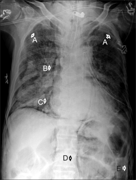
A 74-year-old male patient receiving ventilatory support due to aspiration pneumonia was transferred to the OR from the ICU for an emergency operation of the sigmoid colon volvulus. In the ICU, the preoperative vital signs were blood pressure (BP) of 120/70 mmHg, heart rate (HR) of 70 beats/min, and percutaneous oxygen saturation (SpO2) of 98% on a fractional inspired oxygen concentration (FiO2) 0.4. The preoperative chest X-ray revealed diffuse ground glass opacity in middle lower lung suggesting aspiration pneumonia (Fig. 1). The patient had been ventilated mechanically using synchronized intermittent mandatory ventilation mode with a tidal volume of 450 ml at a respiratory rate of 16 breaths/min, inspiratory : expiratory ratio of 1 : 2, positive end-expiratory pressure (PEEP) of 6 cmH2O, and FiO2 of 0.4. The self respiratory rate was 18-21 breaths/min. Arterial blood gas analysis on a FiO2 of 0.4 revealed a pH, PO2, PCO2 and SpO2 of 7.42, 125.9 mmHg, 43 mmHg and 98%, respectively. The patient received manual ventilation with a self-inflated resuscitator bag (Ambu bag) during transport.
Upon entrance into the OR, he suffered cyanosis and showed a delirious mental state. The SpO2 was 64%. Electrocardiogram revealed tachycardia (HR 130 beats/min) and the BP was 100/70 mmHg. The physical examination showed decreased breath sounds on the left. Manual ventilation was difficult and an adequate tidal volume was not delivered, even with considerable effort. Endobronchial intubation was suspected. The ETT was secured at 22 cm from the ETT tip and was found to be patent. It was then withdrawn slowly to 20 cm. However, the breath sounds and SpO2 did not change. At that moment, subcutaneous emphysema with crepitus and hyperresonant percussion on the left abdomen was found. A pneumothorax was strongly suspected. Accordingly, rapid insertion of a 16 gauge angiocatheter into the left side of the chest was performed and the air was released immediately. Immediately, a 28-French chest tube was inserted into the left side of the thorax. The cyanosis disappeared and vital signs were improved. The SpO2 increased but was below 90%. A chest X-ray was taken, which revealed bilateral pneumothorax, pneumomediastinum, pneumopericardium, pneumoretroperitoneum, and subcutaneous emphysema (Fig. 2). A chest drain was also inserted into the right side of the thorax. The SpO2 increased to 100%. The operation was postponed until the patient improved and transferred to the ICU. The sigmoidectomy was performed 10 days later. However, the patient expired because of an aggravation of pneumonia and residual pneumothorax on the 12th postoperative day.
Discussion
The most widely recognized form of barotrauma is the presence of extra-alveolar air. The most common mechanism for extra-alveolar gas formation is a disruption of the alveolar epithelium, leading to the entry of gas into the perivascular sheath. The gas then dissects within this sheath toward the mediastinum where it decompresses through other fascial planes producing the radiographic and clinical manifestations of pneumothorax, subcutaneous emphysema, subpleural air cysts, pneumomediastinum, pneumopericardium and pneumoperitoneum [4]. The risk of barotrauma is increased by excessive peak inspiratory pressure, high PEEP, high transalveolar pressure, alveolar overdistention and underlying lung injury [1]. The incidences of pneumothorax in patients receiving mechanical ventilation varies from 4 to 15% but might be significantly higher in patients with adult respiratory distress syndrome, status asthmaticus, and aspiration pneumonia [5]. In the present case, underlying aspiration pneumonia might have predisposed the patient to the development of barotrauma.
Manual ventilation is frequently required during intrahospital transpor t. Because clinicians are relatively unconstrained in manual ventilation, the ventilatory patterns can differ significantly among clinicians, and may be inappropriate and deleterious in certain circumstances [6]. A high airway pressure with excessive or inappropriate manual ventilation can cause a pneumothorax and/or other pulmonary air leak problems. In this case, it is likely that the inappropriate manual ventilation induced a high airway pressure or alveolar overdistension, resulting in these complications. The accuracy and stability of manual ventilation can contribute to the quality of ventilation and prevent barotrauma. Accurate and stable manual ventilation can be provided by appropriate training and when a manometer is used in conjunction with the bag. Therefore, physicians should train with a test lung in order to help them realize the ventilation patterns they may generate during manual ventilation.
In this case, the possibility of a preoperative pneumothorax that might have developed in the ICU and remained undetected in the preoperative chest X-ray could not be excluded. It is difficult to make a radiographic diagnosis of a pneumothorax on a portable chest X-ray film taken in the ICU [5]. The traditional radiographic hallmark of a pneumothorax may not manifest in portable supine films of patients with a pneumothorax. In addition, concurrent lung disease might lead to different distributions of free air in the pleural space than in patients with relatively normal lungs [7]. Despite the supine chest x-ray film being reviewed carefully, a pneumothorax can be missed without using other imaging modalities or techniques. Several studies reported that computed tomography (CT) scans of the chest may be more sensitive in detecting a pneumothorax in ICU patients than portable chest radiographs [8-10]. However, it is not always practical or safe to transport critically ill patients for a CT scan to exclude a latent pneumothorax, particularly when the patient is hemodynamically unstable. A preoperative CT scan was not performed in this case.
Manual ventilation can shift the secured ETT within the trachea. Therefore, the migration of an already marginally acceptable ETT position by manual ventilation during transport is believed to be the cause of these complications. The ETT tip was sited near the carina in the preoperative chest X-ray. During transport, migration of the ETT tip to the endobronchial position might lead to a high airway pressure that can result in barotrauma. In the present case, the deep placement of an ETT was not detected in the ICU. Auscultation is not sensitive in detecting ETT tip placement in the main bronchus. Sugiyama et al. [11] demonstrated normal breath sounds with endobronchial depths of 1.5-2.0 cm, and did not detect the disappearance of breath sounds until a depth of 3.2 cm. Ezri et al. [12] reported there were no signs of a change in SpO2 in the cases of endobronchial intubation.
Tension pneumothorax is believed to occur when the communication between the lung parenchyma and pleural space acts as a one-way valve, allowing air to enter the pleural cavity during inspiration but trapping air during expiration. The development of tension is dependent on the pressure gradient between the intrapleural pressure and alveolar pressure. Ventilation will increase the gas flow through the pleural defect, allowing more air to pass per unit time and a more rapid increase in intrapleural pressure with earlier mechanical compressive effects and rapid progress to cardiorespiratory collapse. Therefore, tension pneumothorax develops more commonly in patients on mechanical ventilation than in spontaneously breathing patients. Moreover, ventilated patients with tension pneumothorax deteriorate much more rapidly, and sudden cardiac arrest is more likely [13]. The immediate treatment for tension pneumothorax is needle decompression, which is only a temporary measure until tube thoracostomy can be performed. The definitive treatment of tension pneumothorax is tube thoracostomy.
Strange [14] reported that a clinical diagnosis could be made immediately in 61% of patients with a tension pneumothorax, of which 7% had died. A mortality of 31% was recorded in a group of patients in which clinical signs preceded the diagnosis by 30 minutes to 8 hours. Tension pneumothorax occurred more frequently in patients with an initially misdiagnosed pneumothorax than in patients with a correctly diagnosed. The risk factors predisposing a patient to a misdiagnosis of a pneumothorax in the ICU include mechanical ventilation required at the time of development of a pneumothorax, atypical radiographic location of the pneumothorax, altered mental status exhibited at the time of pneumothorax presentation and the development of a pneumothorax after peak physician staffing hours. In this case, the development of a pneumothorax was not recognized during transport. The intubation state and transport without a specially trained transport team might have contributed to the failure to make an early diagnosis of a pneumothorax during transfer. Knowledge of the factors predisposing the patient to this problem should allow a higher index of suspicion for a diagnosis of a pneumothorax in critically ill patients and possibly improve the early recognition of a pneumothorax.
In conclusion, inappropriate manual ventilation, deep placement of an ETT and aggravation of an undetected preoperative pneumothorax are likely to have contributed to these complications. Accurate and stable manual ventilation, appropriate placement and thorough management of an ETT, as well as an immediate diagnosis and correct treatment can prevent this outcome. In addition, the ability of continual monitoring and appropriate patient care during transport is essential for preventing transport-related morbidity and mortality.