Suprascapular notch cross-sectional area on MRI is not highly accurate in the diagnosis of suprascapular nerve entrapment: counter point of view
Article information

We read with interest the recently published original article by Park et al. [1] presenting the claim that the suprascapular notch (SSN) cross-sectional area is a highly accurate candidate magnetic resonance imaging (MRI) diagnostic indicator for suprascapular nerve (SN) entrapment. We appreciate the authors’ attempt to explore additional parameters using different MRI methods. However, this study contained vital anatomical errors that need to be addressed and clarified. In addition, the claim that their analysis of the SSN space and correlation to SN entrapment is novel and has not previously been reported is inaccurate. We would like to direct the authors’ attention to the original article published by Al-Redouan et al. [2] and the articles cited in that study. Additionally, the authors’ claims regarding the accuracy of their method, or rather, the efficacy of their approach, were not driven by a supporting power study but rather a qualitative descriptive study with a sample size of 10. Park et al. [1] also did not compare their MRI method with other explored modalities. We kindly refer the authors to the article by Jezierski et al. [3] that elaborated on ultrasound as an imaging modality revealing the influence that the different morphological types of SSN have on visualization.
The study by Park et al. [1] also presented an MRI figure demonstrating the SSN cross-sectional area that included anatomical errors in the localization and bordering of the SSN. This section is in fact more dorsal to the SSN and is within the suprascapular canal (SSC), which resides within the spinoglenoid fossa (SGF) [4]. In Fig. 1, we present a serial frontal section of the shoulder on an MRI of the SSC to clarify the anatomy. Localization of the SSN is achieved by identifying its bordering anatomical landmarks. The base of the coracoid process borders the SSN laterally and can be easily navigated on cross-section (Fig. 1A). The authors mentioned that MRI frontal sections cut through the SSN in rather oblique planes, which we do agree with. Therefore, the medial border forming the second anatomical landmark, which is the medial peak of the SSN where the omohyoid muscle attaches, is poorly visualized on MRI and is not usually accurately seen. The space delineated in the figure presented by Park et al. [1] is a site in the middle of the SSC [4], which corresponds to our Fig. 1D. It is an anatomically visible groove that contains the traveling suprascapular neurovascular bundle that resides within the SGF and is roofed by the supraspinatus muscle. The SGF is identified by its laterally bordering glenoid and medially bordering spinoacromial arch proximal to the spinoglenoid notch (SGN). The spinoacromial arch may not be visible on the same plane as the MRI owing to the obliquity of its trajectory. However, because the SGF connects SSN and SGN [4], this connection can be confirmed by navigating the proceeding sections where the SGN is detected by observing the base of the spine of the scapula between the supraspinatus and infraspinatus muscles (Fig. 1E). In fact, this is a common imaging trap, as described by Podgórski et al. [5] in their ultrasound study, in which they explained that capturing a section behind the SSN yields a pseudo-notch image on ultrasound. This imaging concept is the same on MRI and computed tomography of the SSC.
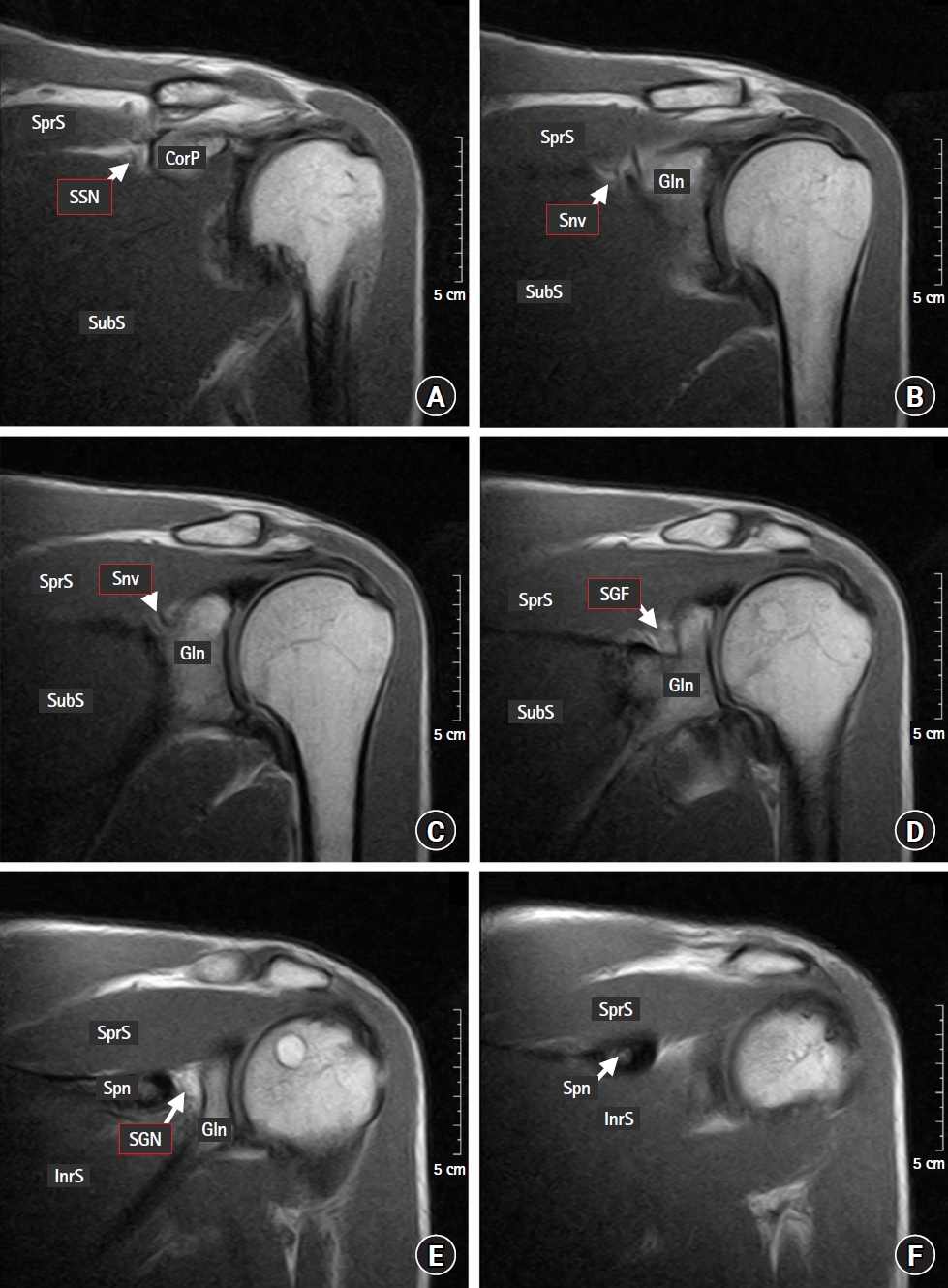
Retrospective MRI frontal section series illustrating the suprascapular notch and suprascapular canal anatomy. SSN: suprascapular notch, Snv: suprascapular neurovascular bundle traveling within the suprascapular canal, SGF: spinoglenoid fossa housing the suprascapular canal, SGN: spinoglenoid notch, CorP: coracoid process (the base of the coracoid process), Gln: glenoid, Spn: spine of scapula, SprS: supraspinatus muscle, InrS: infraspinatus muscle, SubS: subscapularis muscle.
Park et al. [1] claimed novelty in their study and asserted both that the SSN area had not previously been analyzed and that no morphological correlation between SSN typing and SN entrapment had previously been reported. However, a previous study by Al-Redouan et al. [2] demonstrated five morphological SSN stenosis patterns using previously established SSN typing systems and included five parameters from statistically driven correlation analyses of dry bones [2]. To accurately measure the cross-sectional area digitally, an observer must be able to delineate the margins of all bordering parameters. This is a major limitation of MRI and thus comparisons with other modalities such as ultrasound, which allows for plane manipulation, should be undertaken. With MRI, measuring the height or width of the SSN could potentially be more accurate than the cross-sectional area since the chance of having a single parameter fully appearing in the visualization plane is much higher. This is not the case when attempting to capture three collective parameters that are aligned in different spatial orientations. The parameters that govern the cross-sectional area have previously been analyzed, and the height versus upper and mid width were demonstrated to be candidate indicators of SSN stenosis in correlation with SN entrapment [2–4].
In conclusion, the SSN cross-sectional area proposed by Park et al. [1] does not appear to be an accurate approach for estimating SSN stenosis; hence, its reliability as an indicator to assess SN entrapment is questionable. Rather, examining the height and width of the SSN individually could be used to detect SSN stenosis more accurately [2]. MRI is a useful modality for screening the surrounding tissues of the SSC for pathologies [4], while ultrasound has a greater potential for navigating SSC intervals because an observer can manipulate the probe orientation and, thus, the projecting planes [3].
Notes
Funding
This work was supported by The Grant Agency of Charles University (GAUK No. 1720119).
Conflicts of Interest
No potential conflict of interest relevant to this article was reported.
Author Contributions
Azzat Al-Redouan (Conceptualization; Investigation; Project administration; Visualization; Writing – original draft)
David Kachlik (Supervision; Validation; Writing – review & editing)