Cardiac arrest due to intracranial hypotension following pseudohypoxic brain swelling induced by negative suction drainage in a cranioplasty patient: a case report
Article information

Abstract
Pseudohypoxic brain swelling (PHBS) is known to be an uncommon event that may occur during and following an uneventful brain surgery, when negative suction drainage is used. The cerebrospinal fluid loss related to suction drainage can evoke intracranial hypotension that progress to PHBS. The main presentations of PHBS are sudden unexpected circulatory collapses, such as severe bradycardia, hypotension, cardiac arrest, consciousness deterioration and diffuse brain swelling as seen with brain computerized tomography (CT). We present a stuporous 22-year-old patient who underwent cranioplasty under general anesthesia. The entire course of the general anesthesia and operation progressed favorably. However, the time of scalp suture completion, sudden bradycardia and hypotension occurred, followed by cardiac arrest immediately after initiation of subgaleal and epidural suction drainage. After successful resuscitation, the comatose patient was transferred to the neurosurgical intensive care unit and PHBS was confirmed using brain CT.
Extradural and/or subgaleal drainage with a vacuum device is often used in neurosurgery to prevent postoperative hematoma, nevertheless, several cardiovascular complications such as bradycardia, hypotension or even cardiac arrest have been reported to likely be associated with this procedure [1]. Pseudohypoxic brain swelling (PHBS) induced by suction drainage should be considered in patients with unexpected neurological and cardiovascular deteriorations. Intracranial hypotension associated with venous congestion is known to be the main pathomechanism of PHBS [2].
We report a case of a patient having sudden cardiovascular collapse immediately following the initiation of suction drainage in elective cranioplasty. We were able to rule out any common harmful ischemic incidences such as hypotension, hypercapnia or hypoxia during anesthesia. PHBS was confirmed by postoperative brain computerized tomography (CT) after the patient was transferred to the neurosurgical intensive care unit (NICU).
Case Report
A 22-year-old, stuporous, but previously healthy man, 182 cm in height and 70 kg in weight, was admitted to the emergency room with an accidental injury from a fall. An emergent brain CT demonstrated acute traumatic subdural hematoma, thus an emergent decompressive craniectomy and hematoma removal was performed under general anesthesia without any remarkable incidences. After the first operation, the patient had not made favorable progresses, therefore several further operations such as brain abscess drainage, external ventricular drainage catheter insertion, cerebral lobectomy, ventriculoperitoneal shunt were performed during a 4 month period. Finally, the patient had cranioplasty.
The vital signs of the patient scheduled for cranioplasty were a systolic/diastolic blood pressure of 110/70 mmHg, heart rate 104 bpm, respiratory rate via tracheostomy 20 /min and the patient was still in a stuporous state. Preoperative laboratory data were within the normal range, and the chest X-ray revealed improvement of focal pneumonia and an arterial blood gas analysis in the tolerable range (ABGA; pH 7.418/PaCO2 39.5 mmHg/PaO2 104 mmHg with 2 L/min oxygen inhalation). An electrocardiogram (ECG) performed before the surgery showed sinus tachycardia, and transthoracic echocardiography revealed a preserved global left ventricular systolic function (ejection fraction; 69%). The preoperative brain CT sacn showed a marked shrinkage of the left cerebral hemisphere and an increase in ventricular size (Fig. 1). The general anesthesia was induced with 125 mg thiopental sodium and 50 mg rocuronium, without any premedication. The vital signs were monitored using noninvasive blood pressure (NIBP), ECG and a pulse oximeter. The tracheostomy tube was changed for a 7.5 mm cuffed tracheostomy tube. Following the induction of anesthesia, the right dorsalis pedis artery was cannulated with a 22 gauge catheter, and the right femoral vein was cannulated with an 18 gauge catheter. General anesthesia was maintained with balanced anesthesia using oxygen-air-sevoflurane (0.5–1.5%) and supplementary remifentanil (4–6 µg/kg/h) infusion. Cranioplasty was carried out under controlled ventilation, keeping within 10% fluctuation of the vital signs and ETCO2 with intermittent injections of ephedrine, phenylephrine and esmolol. The ABGA was checked immediately after skin incision, and the results were a pH of 7.436, PaCO2 35.1 mmHg, PaO2 219.1 mmHg, and base excess –0.5. Intravenous fluid replacement consisted of 500 ml plasma solution and 50 ml lactated Ringer solution, and the estimated blood loss was 200 ml. After 2 hours of operation, the dura was closed, and the bony fragment was wired into place. Subsequently, drainage catheters were introduced into both the subgaleal and epidural spaces. By the time the scalp sutures were completed, a closed negative suction through the catheters had been applied. Approximately 30 seconds after suction drainage, sudden asystole of ECG, the disappearance of ETCO2 wave and a flat line arterial wave form were noticed. Instantly, the suction drainage was stopped, and we started external cardiac massage for approximately 20 seconds. Two times injections of 500 µg epinephrine were administered under the diagnosis of cardiac arrest. The cardiac rhythm was resumed to 120 bpm, and systolic blood pressure was regained to 150 mmHg immediately. The ABGA was checked after resuscitation, and the results were in the normal range (pH 7.418, PaCO2 39.5 mmHg, PaO2 103.3 mmHg and base excess 0.2). The patient was transferred to the NICU with a controlled AMBU bagging.
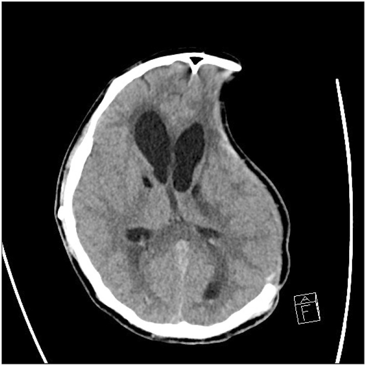
Preoperative brain computerized tomography shows marked shrinkage of left cerebral hemisphere, markedly increased ventricular size and scant amount of intraventricular hemorrhage.
He went into a coma and showed partial spastic movement of extremities, suggesting a sudden increase in intracranial pressure (ICP). Emergent brain CT (Fig. 2) revealed extensive diffuse brain swelling. Due to the fact that there were no specific causes of brain edema during the operation or general anesthesia, the negative vacuum suction drainage was presumed to be the main cause, resulting in the patient being diagnosed with PHBS. The patient received agents to reduce ICP, in addition to general supportive care with controlled ventilation. He improved gradually in the NICU, and reached a stuporous state with self-respiration 3 months postoperatively.

Postoperative brain computerized tomography immediately taken at neurosurgical intensive care unit shows diffuse brain swelling and obliteration of ventricles mimicking diffuse hypoxic brain edema.
Discussion
Van Roost et al. [2] reported PHBS as a newly defined complication after uneventful brain surgery. The main clinical characteristic that appear on brain CT scans include brainstem dysfunction and diffuse brain swelling, especially involving the bilateral basal ganglia and thalami. PHBS has variable prognoses from a fatal state to a full recovery. The main pathomechanism of PHBS is assumed to be a decrease in ICP due to sudden excessive depletion of cerebrospinal fluid (CSF) [3].
An increase in ICP can cause the Cushing response, characterized by the occurrence of hypertension, bradycardia, and respiratory irregularities [4], resulting from the stimulus of pressure on the brainstem [5]. The opposite of the Cushing response, intracranial hypotension, is also though to be a possible cause of circulatory changes such as bradycardia, hypotension and even cardiac arrest [6].
It is known that intracranial hypotension, induced by an extradural suction drainage system for the prevention of postoperative hematoma formation, may occur in neurosurgical procedures. This iatrogenic intracranial hypotension may develop during or after variable neurosurgical procedures when applying negative suction pressure to extradural or epicranial drainage after craniotomy [7], after sudden decompression of hydrocephalus [8], or after spine surgery related to CSF loss during the operation [9]. Because the burr hole trephination and cranial bone incisions are not water proof, negative pressure subgaleal suction may provoke acute loss of the CSF and a decrease in ICP. A rapid fatal case has been reported following a drainage volume of 100-ml CSF. However, a small loss of CSF by gravity can cause PHBS [2]. After a loss of CSF, the brain can expand to fill up free space that was previously filled with gas and fluid including CSF and exsanguinated blood. The pathomechanism of bradycardic rhythm disturbances following intracranial hypotension was reported as a consequence of rostral migration of the brain or brainstem. The pressure changes of the brainstem and hypothalamus can be caused by alteration of sympathetic outflow to induce lethal bradycardia, hypotension, or both [10].
In the present case, firstly, there were no specific events during the entire anesthesia or operation, however, sudden severe bradycardia and cardiac standstill were noticed immediately after initiation of suction drainage through the subgaleal and epidural catheter. The exact amount of CSF was not calculated. Secondly, the postoperative brain CT scan showed diffuse brain swelling. Consequently, we were able to confirm PHBS in this patient. This patient already had shrunken brain hemispheres and a widened intraventricular space, and the negative suction pressure increased the free space after cranioplasty and could evoke a more diffuse brain swelling. We could postulate that this was the reason why severe PHBS happened in this case.
In conclusion, unexpected circulatory collapses such as hypotension, severe bradycardia, and cardiac arrest can develop following intracranial hypotension due to loss of CSF through the negative suction drainage in variable neurosurgical procedures. Anesthesiologists and neurosurgeons should consider, be alert, and cope with potential perioperative circulatory collapses or clinical deteriorations related to PHBS following intracranial hypotension in uneventful brain surgery.