Aseptic subcutaneous inflammation presenting as late onset back pain after uneventful epidural anesthesia
Article information

We encountered a case of aseptic subcutaneous inflammation following epidural anesthesia (EA), which we would like to share with the journal.
A healthy woman (26 years of age; G1P0) was admitted in active labor following uneventful pregnancy. She sought EA for labor pain. Antiseptic cleaning was performed using a chlorhexidine stick, and the target area was draped aseptically. After allowing an adequate time to dry, the skin and underlying subcutaneous tissues at the target needle-entry site paramedian to the L3–4 intervertebral space were infiltrated using 2% lidocaine. An 18-gauge epidural set was inserted in the epidural space to locate it using the loss of resistance to saline technique. The epidural space was uneventfully identified at a depth of 5 cm on the first attempt. Subsequently, the epidural catheter was threaded, and 5 cm of the epidural catheter was retained in the epidural space. After a negative result by the test dose, a loading dose of levobupivacaine was administered. Subsequently, an epidural infusion was started based on our institutional protocol. The patient had instant pain relief and remained comfortable throughout.
Ultimately, a caesarean section was required because of the failure to progress, and it was successfully performed using an epidural “top-up.” At the end of the surgery, the epidural catheter was removed, and a sterile dressing was applied. The total duration of epidural catheter insertion was about 4.5 hours. She received standard antibiotic prophylaxis and postoperative care. Her recovery was uneventful, and she was discharged after 2 days.
She returned on the 7th postoperative day with new-onset atypical back pain without a clear dermatomal pattern. She complained of exquisite pain at the needle-entry site radiating to the mid-thoracic region, contrary to a downward radiation, without any radicular involvement. She did not have fever, headache, or signs of meningism, except slight erythema at the needle-entry site. Her C-reactive protein (CRP) level and white blood cell count remained normal. Her neurological examination was unremarkable.
She was treated conservatively with analgesics, antibiotics, and physiotherapy. As severe pain persisted, interfering with childcare, neurology consultation was arranged, which did not reveal additional information. Magnetic resonance imaging (MRI) revealed inflammation of the subcutaneous tissues overlying the L1–5 vertebrae (Fig. 1). As there was no evidence of drainable fluid or hematoma, no further intervention was advocated other than continuing the conservative management. Her pain reduced steadily, and she recovered fully after 5 days.
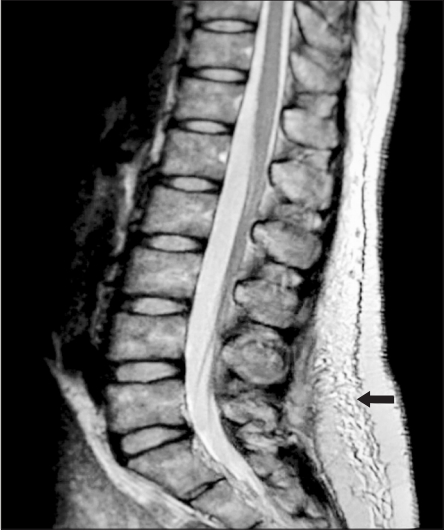
Magnetic resonance imaging shows inflammation in the deep subcutaneous tissues, which are superficial to the paraspinal musculature in the midline, overlying the L1–5 vertebrae. The arrow is pointing to the area of inflammation.
Infective complications after EA can occur because of microbial infection during the procedure, through the needle-puncture site, or seeding of microorganisms from the blood after the traumatic procedure [1]. Non-infectious complications in and outside the neuraxis are also reported after EA [2,3]. Chlorhexidine, which is used for skin asepsis, is cytotoxic and neurotoxic [4,5], but chlorhexidine-induced aseptic inflammation of subcutaneous tissues outside the neuraxis presenting as differential diagnosis after uneventful EA has not been reported. The atypical clinical presentation and MRI findings baffled us. Lack of signs of infection (fever, leukocytosis, raised CRP) or drainable fluid excluded the diagnosis of infection. The only positive finding was erythema at the needle-entry site and MRI suggestive of possible inflammation. After multidisciplinary discussions, we suspected aseptic inflammation caused by inadvertent entry of cytotoxic chlorhexidine as the most probable diagnosis. Chlorhexidine may not have completely dried before administering EA. This probably led to the entry of cytotoxic chlorhexidine causing tissue inflammation. Our assumption was proven by uneventful recovery of the complication using conservative management.
Non-infective subcutaneous inflammation outside the neuraxis presenting as late-onset atypical back pain has not been reported previously. Therefore, we would like to create awareness among fellow practitioners of this atypical presentation as a potential differential diagnosis and to reassure them of its uneventful resolution.
Notes
Conflicts of Interest
No potential conflict of interest relevant to this article was reported.
Author Contributions
Pradipta Bhakta (Conceptualization; Data curation; Methodology; Supervision; Writing–original draft; Writing–review & editing)
Brian O’Brien (Conceptualization; Methodology; Writing–review & editing)
John Richard McNamara (Conceptualization; Data curation; Writing–review & editing)