The prone position provides an excellent environment for surgical approach to the spine and dorsal anatomy. This position, however, can cause hemodynamic changes, such as decrease in arterial blood pressure, mainly due to decreases in stroke volume, cardiac index, and venous return [1-5]. This position also increases intra-abdominal pressure causing complications, such as abdominal compartment syndrome [4]. The Jackson spinal table(Mizuho ISO, USA) has minimal effects on cardiac function and decompresses the abdomen by allowing it to hang freely [4,5].
However, blood may pool in the splanchnic vessels of the abdomen when the it is allowed to hang freely, thereby decreasing venous return and leading to a reduction in cardiac output and systemic hypotension. This case report describes an obese patient who experienced sudden hypotension and hemodynamic collapse soon after prone positioning on a Jackson spinal table; the patient immediately recovered after the use of an external abdominal support.
Case Report
A 75-year-old female patient (weight 78 kg, height 152 cm, body mass index 33.7 kg/m2) with lumbar spinal stenosis in L1/2 and L2/3 was scheduled for an elective surgery involving posterior lumbar spinal fusion and posterolateral interbody fusion. Her previous medical history included diabetes mellitus and Parkinson’s disease. She had been on insulin and levodopa. She had no abnormalities in pre-operative evaluations.
General anesthesia was induced using intravenous propofol, remifentanil and rocuronium. After an uneventful endotracheal intubation, a left radial arterial catheter and a right internal jugular venous catheter were placed. Anesthesia was maintained using desflurane 6 vol% and remifentanil infusion at 0.1 μg/kg/min. The vital signs were as follows: arterial blood pressure, 107/69 mmHg; heart rate, 100 beats/min; respiration rate, 12 breaths/min; and peripheral capillary oxygen saturation (SpO2), 100%.
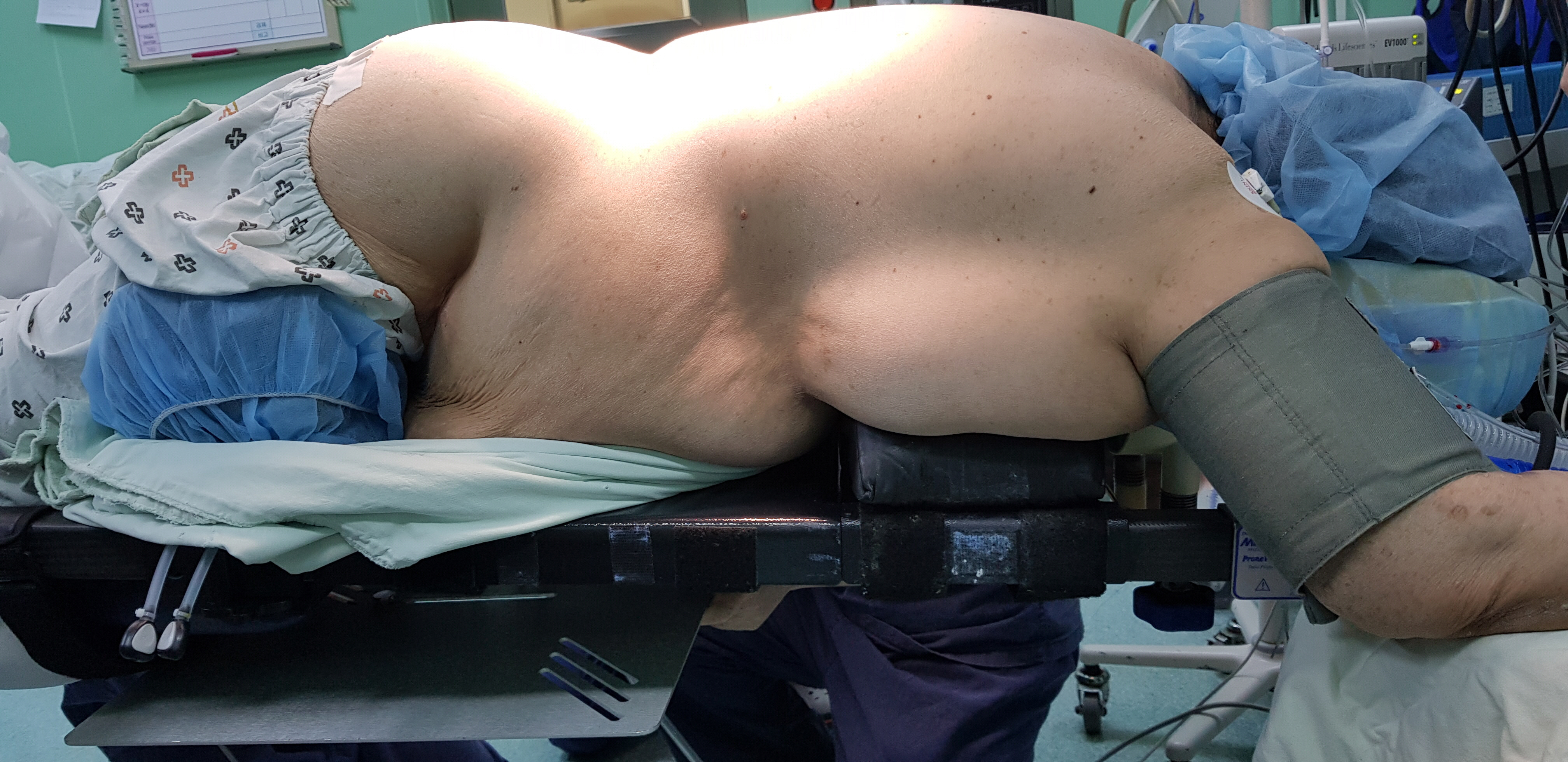
Before the surgery, the patient was positioned in the prone position on a Jackson spinal table. Her abdomen was allowed to hang freely, and severe sagging of the abdomen was observed (Fig. 1). Upon positioning, her arterial blood pressure suddenly dropped to 45/35 mmHg, and the arterial waveform soon disappeared. Heart rate increased by less than 10%, while a normal sinus rhythm was maintained on the electrocardiogram, and SpO2 was 100%. End-tidal carbon dioxide, airway pressure, and body temperature remained unchanged.
The hemodynamic collapse was refractory to preload challenge using boluses of crystalloids. There was also no hemodynamic response to repetitive, dose-increasing boluses of ephedrine, phenylephrine, and even epinephrine. We suspected that these changes were due to mechanical causes that which might have disturbed her normal blood circulation and decided to lift her hanging sagged abdomen. When an external abdominal support was placed (Fig. 2), the arterial waveform and vital signs were rapidly restored.
Once hemodynamic stability was achieved, the surgery began and lasted for 6 hours uneventfully. Before changing her to the supine position, we reproduced the problematic situation by removing the external abdominal support; the abdomen hung freely again, and the sudden drop in arterial blood pressure and the disappearance of arterial wave reappeared. These events quickly disappeared when the external abdominal support was applied. The patient was returned to the supine position and extubated without any complications, including an increase in intra-abdominal pressure and development of abdominal compartment syndrome. She was admitted in the surgical intensive care unit for a day, and there was no other complication until discharge. The written informed consent for the use of images and details of the case for publication of this report has been obtained.
Discussion
Hypotension is a common complication observed during anesthesia and surgery. Common risk factors of intra-operative hypotension are deep anesthesia, hypovolemia, hemorrhage, heart failure and pulmonary embolism.
The prone position is used for a variety of procedures and surgeries, and hypotension is a commonly encountered complication. Several studies in the literature focused on physiologic changes, other than the above-mentioned risk factors, that result in hypotension when an individual is in the prone position. This position causes hypotension by decreasing stroke volume and cardiac index [1]. A decrease in preload is considered responsible for a reduction in stroke volume. Preload can be decreased by blood sequestration in dependent parts of the body, aortocaval compression, increased intrathoracic pressure with poor positioning and chest wall compression, and positive-pressure ventilation [2]. Significant pelvic and abdominal compressions cause an increase in intraabdominal pressure or direct compression of the inferior vena cava that leads to venous pooling and decreased venous return [3]. The increase in thoracic pressure reduces left ventricular compliance and filling, resulting in decreased ventricular volume, stroke volume, and cardiac index [4].
Among several prone positioning systems, the Jackson spinal table produces minimal effects on cardiac function, including the cardiac index and stroke volume [5]. Moreover, the table allows the abdomen to hang freely taht decreases the pressure on the abdomen, especially in obese patients. The external abdominal support is not usually recommended because it can cause changes in the compliance of the abdomen [6].
Sudden hemodynamic collapse is a rare complication in surgery in the prone position. Several cases of hemodynamic collapse manifesting sudden, profound, and refractory hypotension have been reported. Patients with anatomic deformities, such as pectus excavatum or Marfan’s-like features that could compress the thoracic cavity, developed intraoperative refractory hypotension [7-9]. In cases of scoliosis, mechanical compressions on the mediastinum and chest that led to loss of cardiac function and hemodynamic collapse, showing a feature of obstructive cardiogenic shock, were also reported [10,11]. These mechanical compressions were caused by anatomical deformities.
In this case, the patient had no anatomical deformities. Hypovolemia, decreased vascular resistance, and decreased cardiac contractility were ruled out by fluid challenge and the use of vasopressors and inotropes. Hypoxia did not occur and other vital signs and monitoring parameters including end-tidal carbon dioxide, airway pressure, and body temperature remained unchanged. All possible factors were ruled out, excluding the intraabdominal mechanical causes that inhibit venous return to the heart. Her severely sagged abdomen seemed to cause the sudden hemodynamic collapse. We suspected that this abdominal sagging pulled her abdominal wall and intraabdominal contents downwards, thus, leading to kinking of the inferior vena cava or blood sequestration in the abdomen. Subsequent sudden obstruction of inflow to the heart was then suspected to have caused the hemodynamic collapse. The hemodynamic instability was simply resolved by applying an external abdominal support, and it seemed to restore venous return.
Factors that increase intrathoracic or intraabdominal pressure cause hypotension and hemodynamic collapse. In obese patients, the distended abdomen can be compressed in the prone position, severely enough to increase intraabdominal pressure that can cause circulatory collapse with or without an abdominal compartment syndrome. The Jackson spinal table is widely used to prevent these complications. When the patient is placed on this table, less pressure is exerted on the abdomen, and the risk of increasing abdominal pressure and development of abdominal compartment syndrome is reduced.
However, in obese patients like the patient in this case, using the Jackson spinal table can cause kinking of a large vessel or severe blood sequestration in the abdomen by hanging the abdomen and pulling intraabdominal contents downwards. Although the application of an external abdominal support is not usually recommended in the prone position, this case showed that in some situations it may reduce the risk of hemodynamic instability. If hemodynamic collapse occurs with prone positioning, the application of an external abdominal support to lift up the hanging abdomen should be considered, and careful observation of the intraabdominal pressure is required.