Pneumothorax in a post-anesthetic care unit after right thyroidectomy with left neck dissection -A case report-
Article information
Abstract
A 46-year-old woman underwent a right thyroidectomy with left neck dissection under general anesthesia. The operation was performed successfully for over the course of 3 hours 30 minutes. After extubation, the patient was transferred to post-anesthetic care unit (PACU). After 10 minutes, dyspnea, chest discomfort, desaturation was suddenly occurred. Intubation was performed in PACU. The emergency chest X-ray revealed a right pneumothorax, and the patient was treated by chest tube insertion. The patient was improved and was discharged uneventfully from hospital 8 days later.
Pneumothorax is a complication that can occur during general anesthesia, mid- or post-surgery. It can occur from thoracic damage or alveolar ruptures due to high positive airway pressure [1]. It can also lead to damage of the neck, trachea, throat, esophageal wall, and the abdominal wall, although this is infrequent [2].
With pneumothorax follows thoracic pain, a decline of oxygen saturation, decreased sound through the stethoscope, tachycardia, and reduction in blood pressure. However, these symptoms do not occur under general anesthesia or they are interpreted as physiological changes caused by general anesthesia. If tension pneumothorax progresses due to positive pressure, the patient can fall into a critical state. Thus, it is important to diagnose and treat pneumothorax early. However, it is difficult to diagnose and find the cause of pneumothorax that is caused by surgery that does not entail thoracic damage. There have been many case reports that gas of excessive pressure infused in endoscopic surgery cause pneumothorax [3]. However, pneumothorax occurring in cranio-cervical surgery has rarely been reported. This report focuses on a case where a patient experienced pneumothorax while in the recovery room after a right thyroidectomy with left neck dissection.
Case Report
A 46-year-old female patient, who had had a left thyroidectomy 20 years earler, was admitted for a right thyroid lump that was found during a regular check-up. The neck computed tomography showed that the left lobe of the thyroid gland had been removed during the previous surgery, and the lower right lobe had a 7 mm node. On the left side of the neck, the enhanced image of a 1.5 × 0.9 cm sized lymph node was discovered. In a fine needle aspiration biopsy of the thyroid, papillary thyroid cancer was diagnosed. Thus, the patient was admitted for a right thyroidectomy with left neck dissection. The patient had been taking thyroid hormone pills (Levothyroxine Na 0.1 mg). The blood test before the surgery was within normal range. The chest X-ray and the electrocardiogram (EKG) showed no strange findings. As preanesthetic medication, glycopyrrolate 0.2 mg and butorphanol 0.5 mg were intramuscularly injected 30 minutes before surgery. On the operating room (OR) table, the noninvasive blood pressure monitor, the EKG, the pulse-oximeter, and the capnometer were attached for patient observation.
Upon arriving into the OR, the patient's blood pressure was 120/80 mmHg, her pulse rate was 78 beats pre minute (bpm), and oxygen saturation was 100%. For anesthetic induction, propofol 100 mg and rocuronium 50 mg were intravenously administered. After we induced loss of consciousness and adequate muscle relaxation, we performed an endotracheal intubation with a 7.0 mm tube. There was no complication with intubation. Immediately after intubation, we checked the sound of the patient's respiration. The tube was then set at a depth of 22 cm. Mechanical ventilation was set with the tidal volume at 10 ml/kg and respiration rate at 10 bpm. Anesthesia was maintained by oxygen 2.5 L/min, medical air 2.5 L/min, remifentanil 0.15 µg/kg/min, sevoflurane 1.0-1.5 vol%, and atracurium 7.5 µg/kg/min.
Two and a half hours into the surgery, the blood pressure which was initially set at 130-120/85-65 mmHg momentarily dropped to 85/45 mmHg. The pulse rate was 80 bpm, and oxygen saturation was stable at 100%. When ephedrine 5 mg was intravenously administered and crystalloid solution 300 ml was quickly infused, the blood pressure recovered to 115/65 mmHg and was maintained until the end of the surgery without complications. The vital signs were within the normal range for 3 hours 30 minutes of the surgery. The end-tidal carbon dioxide tension was maintained at 32-35 mmHg, and the airway pressure was 19 mmHg.
After the surgery, 100% oxygen was administered. To induce spontaneous respiration, glycopyrrolate 0.2 mg and pyridostigmine 10 mg were intravenously administered. After confirming a reverse of muscle relaxant, the patient was extubated. After extubation, the pulse-oxygen saturation was maintained at 100% and the patient fully recovered consciousness. The patient did not complain of any pain or discomfort. She was then moved to the recovery room.
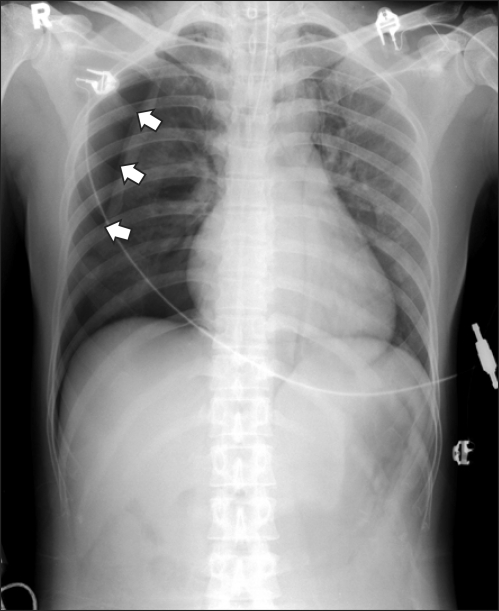
After being moved to the recovery room, oxygen 3 L/min was administered by nasal cannula and the patient's recovery was checked. The vital signs were within the normal range with the blood pressure at 145/100 mmHg, pulse rate 80 bpm, and oxygen saturation 100%. But within 10 minutes of the patient being moved to the recovery room, she suddenly complained of chest pain. Pulse oxygen-saturation had suddenly dropped to 90%. We immediately switched to using the partial rebreathing mask. 100% oxygen 10 L/min was administered. The patient was reassured and made to breathe deeply. The pulse oxygen saturation stabilized at 85%. Stethoscopy of both sides of the thorax revealed that sound of respiration of the right side was considerably faint. Pneumothorax and damage to the right recurrent laryngeal nerve, which commonly occurs in thyroid surgeries, were suspected. Thus, emergency tracheal intubation was prepared. Upon 20 minutes of the patient being moved to the recovery room, she again complained of chest pain and her pulse oxygen saturation had suddenly dropped to 65%. Her blood pressure had reduced to 110/70 mmHg and her pulse rate increased to 100 bpm. By using Mapleson F circuit, manual ventilation was performed. When respiration through both sides of the thorax was heard through the stethoscope, we found that right side of the thorax emitted no sound of respiration. Pneumothorax was strongly suspected. To remedy the pulse oxygen saturation and to stabilize airway maintenance, propofol 60 mg and succinylcholine 60 mg were intravenously administered for emergency endotracheal intubation. After inducing loss of consciousness and adequate muscle relaxation, endotracheal intubation with a 7.0 mm tube was performed. When manual ventilation was completed with 100% oxygen, the pulse oxygen saturation rate elevated to 98%. The emergency arterial blood gas analysis (FiO2: 1.0) showed pH 7.26, pCO2 61 mmHg, pO2 280 mmHg, and SpO2 100%. The chest X-ray taken in the recovery room found a collapsed right lung and pneumothorax (Fig. 1). Forty-five minutes after being moved to the recovery room, the patient's vital signs stabilized. She was manually ventilated and moved to the intensive care unit (ICU). In the ICU, her blood pressure was 142/69 mmHg, pulse rate 105 bpm, and SpO2 94%. Tube thoracostomy was conducted and the outflow of a large amount of air was observed. After the initial outflow, there was no more air outflow. The chest X-ray showed the recovery of the collapsed right lung and extinction of pneumothorax. Three hours after tube thoracostomy, the arterial blood gas analysis (FiO2: 0.4) was pH 7.40, pCO2 41 mmHg, pO2 88 mmHg, and SpO2 97%.

Chest radiograph after re-intubation of endotracheal tube. This shows pneumothorax in right hemithorax (arrow) and partially collapsed right lung.
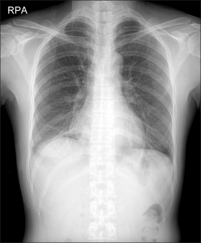
Post-surgery day-2 at 07:00 a.m. the patient was extubated carefully. Her vital signs were within normal range. She was then moved to the general ward. On post-surgery day-5, the thoracostomy tube was removed. On day-8, she was released from the hospital with required ambulant treatment. The chest X-ray on the day of release is shown in Fig. 2.
Discussion
Maclntyre [4] divided the causes of pneumothorax related to general anesthesia into 4 groups. In Group 1, the alveolar rupture causes the air around the sheath near the vessel to flow out of the mediastinum, diffuse in the pleural cavity, and cause pneumothorax. Group 2 involves rupture of the mediastinal pleura that follows damage to the fascial layer with the mediastinal emphysema. In Group 3, the peripheral airway and the pleura are connected directly. Type A is from the alveolar rupture on the surface of the lung without any damage to the pleura. Type B is when the chest wall and the pleura both have ruptures due to damage of the chest wall. It is also when there is damage to the lung parenchyma. Group 4 involves damage only to the chest wall. The internal and external part of the thorax is usually punctured.
The reported causes for general anesthesia-related pneumothorax usually belong to Groups 1 and 4 [1-3]. Alveolar ruptures are caused by excessive positive airway pressure. They occur when pressure increases in the alveoli over a steady period of time rather than in a sudden increase in pressure. In general, if transpulmonary pressure does not go beyond 30-35 cmH2O, alveolar ruptures almost never occur [5]. Additionally, as long as tidal volume during mechanical ventilation does not go below 10 ml/kg and the plateau pressure does not exceed 25-30 cmH2O, lung damage due to pulmonary overinflation almost never occurs [6]. Pepe et al. [7] stated that positive end-expiratory pressure of the usual positive pressure does not increase the rate of occurrence of pneumothorax. Kirby et al. [8] reported that high positive end-expiratory pressure above 18 mmHg increases the rate of occurrence. In the presented case, there were no strange findings in the patient's chest X-ray. She had no past history of any respiratory disease. Because there was no peripheral vascular oxygen saturation decline or hypoxia during the surgery, it is believed there is little chance that the pneumothorax was due to the damage of the airway or of the larynx during the anesthesia. During volume control ventilation with the tidal volume at 10 ml/kg and respiratory rate at 10 bpm, the airway pressure stabilized below 19 mmHg, and positive end-expiratory pressure was not performed. Thus, the likelihood of alveolar rupture due to alveolar overinflation is also low. Therefore, we assume that the pneumothorax occurred from operative damage to the neck and chest or from anatomical causes. We believe that the presented case belongs to Martin et al.'s Group 2.
The most common complication following total thyroidectomy is damage to the recurrent laryngeal nerve [9]. Other complications include hypocalcaemia, hematoma, and inflammation. In general, thyroid surgeries and pneumothorax are not considered to have any cause-and-effect relationship [10]. However, the possibility of pneumothorax occuring during thyroid surgeries is often mentioned. Fewins et al. [11] proposed the hypothesis that the surgery site expands to the pleura during difficult surgeries of radical neck dissection, and pneumothorax occurs. He also hypothesized that air enters through the fascial layer of the surgical site and diffuses into the thorax from the pressure difference by negative pressure. There are some reported cases that suspect pneumothorax is related to thyroid surgeries. Slater and Inabnet [9] reported that 1 of the 2 cases of pneumothorax that occurred after a parathyroid gland surgery was due to alveolar ruptures. In the other case, the parathyroid gland was located near the thymus gland, and in the middle of dissection, there was pleura damage which caused pneumothorax. Taniguchi et al. [12] reported on a patient, who had no strange respiratory findings before surgery, but developed pneumothorax after the thyroid surgery. The patient with thyroidectomy and radical neck dissection had no clear signs of mid-surgery thoracic damage. However, after surgery, air entered through the thinned neck wall by intense respiratory effort. These two factors are assumed to cause the bilateral pneumothorax. In the presented case, because there were no clear signs of chest damage, there is the possibility that air entered through the fascial layer or that by the negative pressure, the air entered through the thinned fascial layer due to the surgery. However, we cannot exclude the possibility that the pneumothorax may have been caused by mid-surgery damage of which we were unaware. Menegaux et al. [13] also reported that re-surgeries of the parathyroid gland increase the possibility of anatomical damage to such places as the recurrent laryngeal nerve. The patient of the presented case had a previous thyroidectomy, so there were severe adhesions in the surgical site. Considering that surgical maneuvering was difficult, there may have been thoracic damage of which the surgical team was unaware.
In spontaneously breathing patients, small pneumothorax can be naturally absorbed and is a minor problem. However, when intermittent positive pressure ventilation is performed for a patient under general anesthesia, the intrapleural pressure suddenly increases and can cause it to develop into tension pneumothorax. Symptoms of pneumothorax in general anesthesia are firstly tachycardia and hypotension, and then hypercapnia and hypoxia. When pneumothrax progresses, it can lead to cyanosis. When tension pneumothorax progresses, it presses the lung of the same side and results in atelectasis. If pneumothorax further progresses, it causes mediastinum displacement, compresses the opposite lung, compresses the vena cava, reduces cardiac output, and triggers rapid circulation failure [14]. Conscious patients experience tachycardia, dyspnea, and chest pain, which can reduce depending on the body position. If pneumothorax further progresses, the patient experiences cyanosis and severe tachycardia, which can lead to loss of consciousness and lung collapse. In spontaneously breathing patients, 10-20% of the progression of pneumothoraces can be observed. Beyond 20%, tube thoracostomy must be done. Patients under mechanical ventilation must be immediately inserted with tube thoracostomy regardless of the size of the pneumothorax [15]. The patient of the presented case was alert after the surgery and breathed spontaneously, when sudden chest pain and decline in peripheral vascular oxygen saturation occurred. At the same time, through use of the stethoscope we found that the sound of the patient's respiration had decreased. Thus, pneumothorax was suspected and confirmed by a chest x-ray. Therefore a tube thoracostomy was performed.
We experienced that pneumothorax can occur infrequently even in neck surgeries without invasive maneuvers such as central venous catheterization, as in the presented case. Pneumothorax is commonly known to occur from laparoscopic surgery. However, an unknown cause during a surgical maneuver must also be suspected if the patient shows hypoxia and tachycardia.
