Bear maul injuries are a rare cause of facial trauma and can cause severe deep lacerations involving the eyes, lips, nose, and maxillofacial structures. These facial injuries require multiple reconstructive surgeries to achieve long-term aesthetic and functional outcomes.
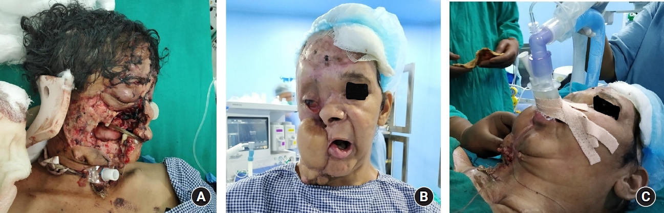
Case scenario 1: A 45-year-old woman (weight: 53 kg, height: 168 cm) presented to the emergency room with a history of an offensive attack on face by a sloth bear. The patient provided informed written consent for the publication of her case report in the medical literature. At the time of presentation, she had facial disfigurement with a right orbital fracture, loss of the right cheek, split palate, and mandibular fracture (Fig. 1A). A tracheostomy was performed owing to difficult airway at the time of presentation, followed by initial debridement and suturing. Seven days later, a free anterolateral thigh flap reconstruction was performed. The flap failed on the 8th post-surgery day because of sudden extensive hemorrhage. A radial artery forearm extracorporeal flap used as salvage partially covered the defect. Finally, a trapezius myocutaneous flap was used for resurfacing, followed by multiple scalp rotation flaps. All five reconstructive surgeries were performed under general anesthesia via the tracheostomy port. Her tracheostomy site was closed 9 months after the injury.
Case scenario 2: The same patient presented for flap revision after undergoing multiple surgeries; we noted overgrowth of the myocutaneous flap over the right cheek and neck (Fig. 1B). Flap revision and cutting were required to enable her to extend her face and open her mouth to achieve aesthetic and functional improvement. An airway examination revealed a lateral mouth opening of only 2 cm, limited neck movement (both flexion and extension), a sterno-mental distance of 5 cm, a thyro-mental distance of 2.5 cm, a patent left nostril, and extensive flap overgrowth over the right side of the face (Fig. 1B).
We prepared our patient with lidocaine nebulization, mouth gargles, and anti-sialagogue 0.2 mg of glycopyrrolate. Inside the operating theater, after standard monitors were attached, pre and para-oxygenation was provided via a nasal cannula. The patient maintained spontaneous ventilation, and aliquots of 1 mg of midazolam and 60 mg of ketamine were given. Mask placement was difficult but was achieved using the two-handed two-operator technique with a mask overlying the left side of the nasal cavity and mouth and the reservoir bag held by the other person. One hundred percent oxygen and sevoflurane was titrated to maintain a minimum alveolar concentration induction of 1.2 under spontaneous ventilation, an i-gel┬« supraglottic airway(┬® Intersurgical Ltd, UK.) size 2.5 was inserted laterally into the mouth by a senior anesthesiologist (Fig. 1C) and placement confirmed by end tidal carbon dioxide and chest rise. The available 2-cm mouth opening could accommodate only an i-gel size 2.5, and it was secured properly and connected to a closed circuit. She was kept on ventilation with a pressure support of 10 cmH2O, partial end expiratory pressure of 5 cmH2O, trigger at 1 L/min, and apnea frequency of 10 /min. On these settings, the patient was generating a tidal volume of 380ŌĆō400 ml, spontaneous breath efforts of 14ŌĆō15 /min, a minimal leak of 20ŌĆō40 ml, peak airway pressure of 14ŌĆō16 cmH2O, and end tidal carbon dioxide of 31ŌĆō35 mmHg. The entire surgery (extraoral) lasted for 45 min with minimal blood loss on spontaneous ventilation with pressure support. The i-gel was removed at the end of the surgery and the patient was shifted to the postoperative area. We had access to a backup tracheostomy and fiberoptic bronchoscope, but it was not working properly. The only other option was to cancel the surgery and awaken the patient. The plans were discussed with the surgeons, patient, and her relatives.
A bear is a strong wild animal that is potentially dangerous and can cause unpredictable serious injuries over the face. In this case, on first arrival in hospital, owing to complete facial disfigurement, a tracheostomy was the only available option; anesthesia for the rest of the facial reconstructive surgeries was administered via the tracheostomy port. In the second case scenario, she underwent closure of the tracheostomy site placed for the anticipated difficult airway owing to the decreased mouth opening, limited neck movement, and no nostril. We had limited options for securing the airway in this case. We prepared the airway with lidocaine gargles, nebulization, and mouth spray, with due consideration taken, not to exceed local anesthetic toxic dosage. We did not have a functional fiberoptic device, and intubation via the same can be tricky in such cases owing to the availability of a single nostril and distorted airway anatomy. Direct laryngoscopy was impossible owing to limited mouth space. We maintained para-oxygenation via a nasal cannula and the patientŌĆÖs ventilation remained spontaneous throughout the surgery. Mask ventilation was difficult owing to the nasal flap fistula and overgrowth over the right side. We meticulously kept the mask over left side of the face and held it opposite the fistula site and inserted gauze pieces in the open areas. We inserted an i-gel under lidocaine preparation of the airway and sedation with midazolam, ketamine, and sevoflurane. The surgeons were ready to perform a tracheostomy in case of difficulty.
We reiterate that not all difficult airways require intubation, although the standard of care in most difficult airway cases is awake fiberoptic intubation. If a fiberoptic device is not available, an i-gel can be used as a definitive device for short procedures to defer tracheostomy and reduce undue patient morbidity. Meticulous preparation of the airway, maintaining spontaneous ventilation, sedation and analgesia maintained by ketamine, and inhalational agent assistance enabled successful i-gel placement without any procedural complications.