Sir, Fracture and migration of central venous catheter is a potentially dreadful complication, which requires early recognition and prompt intervention. Point-of-care ultrasonography (POCUS) is becoming an important tool for early localization and subsequent removal of foreign bodies in the body cavities [1,2]. This technique averts the need for X-ray or computed tomography (CT) imaging, and therefore, the associated radiation exposure. Embolization of fractured venous catheter in the external jugular vein (EJV) has been reported during patient movement, although a valve present at the end of the EJV halts this [3]. Migration of the fragment during the lag period between identification and retrieval causes great difficulty in its removal. POCUS enables real-time detection and correct localization immediately before surgical retrieval.
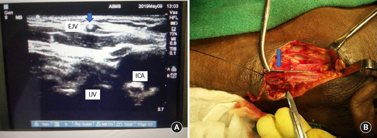
An 84-year-old male patient (written informed consent has been taken from the patient for publication) with diagnosis of pneumonia in the post-chemotherapy phase for acute myeloid leukemia was shifted to the intensive care unit for intermittent non-invasive ventilation. He had a 16-gauge external jugular venous catheter in situ, in view of difficult peripheral venous access. During removal, the catheter broke with the entire sheath inside the vein. Bedside POCUS in the intensive care unit was used to locate the catheter sheath (Fig. 1A). Surgical removal of the broken piece of the catheter was planned. Position of the fragment was re-confirmed by POCUS in the operation theatre just before retrieval, and was removed successfully under local anesthesia and monitored anesthesia care (Fig. 1B). Post-retrieval ultrasound was done to ensure the absence of remnants.
Various scenarios have been reported in relation to fractured intravenous catheters. Doley et al. [4] reported retrieval of a migrated fragment of an implanted central venous catheter through the neck surgically under fluoroscopic monitoring, after its initial detection by X-ray imaging.
A previous study reported fracture of central venous catheter and embolization into heart chambers and subsequently pulmonary vessels due to delayed detection and failure in early retrieval. [5]. Fluoroscopy with X-ray is considered the gold standard in retrieval of fractured catheters [4]. Nonetheless, we found that POCUS could be a suitable alternative for accurate detection and retrieval of broken catheter fragments. Therefore, we present the following advantages of POCUS:
1. Prompt localization of catheter position in case of suspected dislodgement, thus avoiding further displacement.
2. Avoids the need for radiological imaging like CT and shifting of the patient to the CT room, which may be difficult in critically ill patients.
3. Real-time imaging during removal helps to confirm position of the object, and ensures correct site of incision to avoid blind exploration and risk of vascular injury.
4. Confirms complete removal of the object after the procedure
5. Enables locating thrombus (if any), thereby guiding in taking necessary precautions.